Cell Cycle and Cell Division
Cell Cycle and Cell Division PDF Notes, Important Questions and Synopsis
SYNOPSIS
- The sequence of events by which a cell duplicates its genome and synthesises all other cell contents and eventually divides into two daughter cells is called the cell cycle.
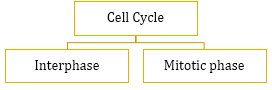
- Interphase involves a series of changes which take place in a newly formed cell and its nucleus before it gets ready for division again. It is also called intermitosis. It is further divided into
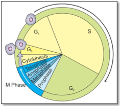
- First gap or G1 phase: Interval phase between mitosis and the initiation of DNA replication. The cell grows to its maximum size to prepare for DNA replication.
- Synthetic or S phase: Synthesis or replication of DNA occurs on the template of the existing DNA strand. The amount of DNA per cell doubles, but there is no change in the chromosome number of the cell.
- Second gap or G2 phase: Synthesis of RNA and proteins continues. Spindle protein synthesis and aster formation take place.
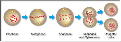
- Phases of Mitosis
|
Phase |
Description |
|
Karyokinesis (Division of the nucleus) |
|
|
Prophase |
|
|
Metaphase
|
|
|
Anaphase
|
|
|
Telophase
|
|
|
Cytokinesis (Division of the cytoplasm) In animals – cell furrow method In plants – cell plate formation |
|
- Phases of Meiosis
- Meiosis-I or Heterotypic Division

|
Phase |
Description |
|
Prophase I |
|
|
|
|
|
|
|
|
|
|
|
|
|
|
|
|
|
|
Cytokinesis (division of the cytoplasm) In animals – cell furrow method In plants – cell plate formation By the end of cytokinesis, each diploid parental cell divides into two daughter cells each with a haploid number of chromosomes but double the content of DNA. |
|
- Meiosis-II or Homotypic Division

- In this division, the two chromatids of each chromosome separate from each other and go to separate daughter cells.
- The number of chromosomes remains the same as produced by meiosis-I.
- Meiosis-II consists of four stages—prophase-II, metaphase-II, anaphase-II and telophase-II.
- Cytokinesis follows karyokinesis of meiosis-II.
- At the end of cytokinesis, two daughter cells are formed, each with half the number of chromosomes and half the amount of nuclear DNA of the parent cell.
Related Chapters
- The Living World
- Biological Classification
- Plant Kingdom
- Animal Kingdom
- Morphology of Flowering Plants
- Anatomy of Flowering Plants
- Structural Organisation in Animals
- Cell : The Unit of Life
- Biomolecules
- Transport in Plants
- Mineral Nutrition
- Photosynthesis in Higher Plants
- Respiration in Plants
- Plant Growth and Development
- Digestion and Absorption
- Breathing and Exchange of Gases
- Body Fluids and Circulation
- Excretory Products and their Elimination
- Locomotion and Movement
- Neural Control and Coordination
- Chemical Coordination and Integration
- Reproduction in Organisms
- Sexual Reproduction in Flowering Plants
- Human Reproduction
- Reproductive Health
- Principles of Inheritance and Variation
- Molecular Basis of Inheritance
- Evolution
- Human Health and Disease
- Strategies for Enhancement in Food Production
- Microbes in Human Welfare
- Biotechnology : Principles and Processes
- Biotechnology and its Applications
- Organisms and Populations
- Ecosystem
- Biodiversity and Conservation
- Environmental Issues