CBSE Class 10 Answered
The intestinal villi are small finger like projections that extend into the lumen of the small intestine. Each villus has many microvilli projecting from its epithelial surface, collectively forming a brush border. Villi are specialised for absorbtion and have very thin walls which are single cell thick. They have a rich blood supply to keep a concentration gradient. Villus capillaries collect amino acids and simple sugars.
Lacteals are lymphatic capillaries found in the villi of the small intestine. They absorb and transport large molecules, fats, and lipids in the digestive system mainly in the form of lipoproteins. The combination of fat and lymph in the lacteals is milky in appearance and is called chyle. This chyle is carried by the lacteals to the lymph vessels in the intestinal wall. Individual lacteals merge to form larger lymphatic vessels that transport the fats to the thoracic duct which empties into the left subclavian vein.
Application Videos
-
Absorption in small intestine
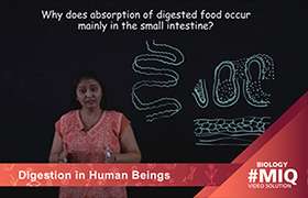 This video explains why absorption of food occurs in the small intestine.
 -
Role of Pancreatic Juice in Digestion
This video explains an assertion reasoning question based on the role of...
-
Human Alimentary Canal

This video explains the parts of the human alimentary canal.
Concept Videos
-
Nutrition in Organisms
 Heterotrophic nutrition and its types; Holozoic nutrition in Amoeba and Hum...
 -
Nutrition in Organisms
 Heterotrophic nutrition and its types; Holozoic nutrition in Amoeba and Hum...
 -
Life Processes
 Life Processes, Nutrition in Animals
 -
Life Processes
 Life Processes, Nutrition in Animals
 -
Life Processes
 Life Processes, Nutrition in Animals