CBSE Class 10 Answered
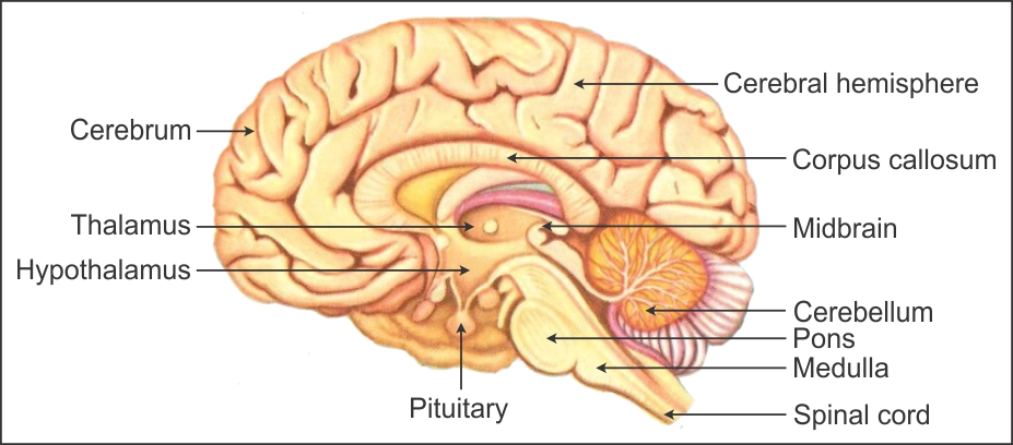
The brain is the main coordinating centre of the body.
|
Part of the brain |
Function |
|
Cerebrum |
The cerebrum is the centre of intelligence, memory, consciousness, will power and voluntary actions. |
|
Thalamus |
The thalamus relays pain and pressure impulses to the cerebrum. |
|
Hypothalamus |
The hypothalamus controls the body temperature and the activity of the pituitary gland. |
|
Midbrain |
This small tube-like part is responsible for reflexes involving the eyes and ears. |
|
Cerebellum |
The cerebellum coordinates muscular activity and balance of the body. |
|
Pons Varolii |
The pons varolii carries impulses from one hemisphere to the other hemisphere and coordinates muscular movements on both sides of the body. |
|
Medulla oblongata |
The medulla oblongata controls the activities of the internal organs, heartbeat and breathing. |
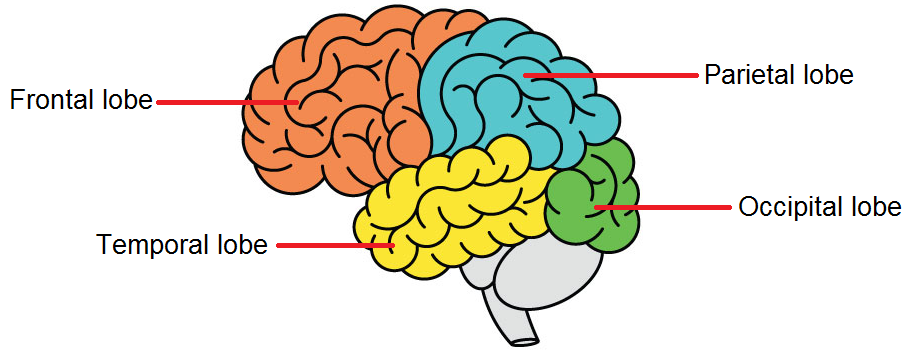
The cerebral cortex is divided into 4 lobes-frontal lobe, temporal lobe, occipital lobe and parietal lobe.
- Frontal lobe – behaviour, intelligence, memory and movement
- Parietal lobe- intelligence, language, reading and sensation
- Temporal lobe- behaviour, hearing, memory, speech, vision
- Occipital lobe- vision
- A kidney is composed of an enormous number of uriniferous tubules.They are also known as nephrons or renal tubules or kidney tubules.
- Nephrons are the structural and functional units of the kidney.
- Each kidney is formed of about 1 million nephrons.
- Nephrons are held together by a connective tissue.
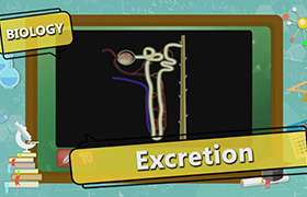
Structure of nephron:
- Each nephron is made of a glomerulus and a renal tubule.
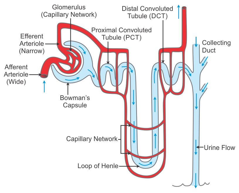
Glomerulus:
- It is a tuft of anastomosing blood capillaries formed by the fine branches of the
afferent arteriole. - These capillaries of the glomerulus again unite to form the efferent arteriole.
- The afferent arteriole is wider than the efferent arteriole.
- The afferent arteriole brings blood into the glomerulus, while the efferent arteriole collects blood from the glomerulus.
Renal Tubule:
The renal tubule comprises the following regions:
Bowman’s Capsule
- The Bowman’s capsule lies in the cortex.
- It is a thin double-walled, cup-like depression.
It is the blind end of the nephron.
- The glomerulus is located in the concave depression of the Bowman’s capsule.
- The Bowman’s capsule and the glomerulus together are called Malpighian Capsule or Renal Capsule.
- The outer layer of the Bowman’s capsule is made of flattened epithelial cells.
- The inner layer is in close contact with the glomerulus and bears special cells called podocytes.
- Podocytes have many feet-like processes called pedicels and minute slit-like pores called slit pores.
Proximal Convoluted Tubule (PCT)
- PCT lies in the cortex.
- It is also known as the first convoluted tubule.
- The Bowman’s capsule continues into the PCT.
- It is lined with a layer of columnar epithelial cells.
- The columnar cells are with numerous microvilli to increase the surface area of absorption.
Loop of Henle
- It lies in the medulla.
- It is U-shaped.
- It is not convoluted.
- It has a descending limb and an ascending limb.
- Each limb has a thick region towards the cortex and a thin region towards the medulla.
- Thick regions are lined with columnar epithelial cells.
- Thin regions are lined with flat epithelial cells.
Distal Convoluted Tubule (DCT)
- It lies in the cortex.
- Its short terminal part is called a collecting tubule.
- The collecting tubule opens into the collecting duct.
- DCT in lined with ciliated columnar epithelial cells.
- The collecting duct receives the contents of many renal tubules.
- The collecting duct is a larger duct which receives collecting tubules of several nephrons.
- Collecting ducts pass into the renal medulla and join with each other to form the ducts of Bellini.
- The ducts of Bellini run through the renal pyramids and open into calyces.
- All calyces open into the pelvis.
- From the pelvis, urine is carried to the urinary bladder through the ureters.
Application Videos


Concept Videos
-
Life Processes
 Life Processes, Excretion
 -
Life Processes
 Life Processes, Excretion
 -
Life Processes
 Life Processes, Excretion