Class 10 SELINA Solutions Biology Chapter 8 - The Circulatory System
The Circulatory System Exercise Ex. 1
Solution A.1
(c) Erythrocytes
Solution A.2
(c) Spleen
Solution A.3
(c) Phagocytosis
Solution A.4
(b) Renal artery
Solution A.5
(a) Carbaminohaemoglobin
Solution A.6
(b) Antitoxins
Solution A.7
(c) Pericardium
Solution A.8
(a) Capillary
Solution A.9
(d) Hepatic portal vein
Solution A.10
(d) Thrombocytes
Solution B.1
(a) Blood platelets - Blood coagulation
(b) Neutrophils - Phagocytosis
(c) Erythrocytes - Transportation of gases
(d) Lymphocytes - Produce antibodies
(e) Bone marrow - Destruction of old and weak RBCs / Production of RBCs and WBCs
Solution B.2
(a) Heart, blood, and blood vessels
(b) Erythrocytes, leukocytes and thrombocytes
(c) Arteries, veins and capillaries
(d) Blood, tissue fluid and lymph
(e) Synovial fluid, urine and vitreous humour
(f) Spleen and tonsils
(g) Plasma and cellular elements
(h) Open circulatory system and closed circulatory system
(i) Haemin and globin
(j) Pulmonary circulation and systemic circulation
Solution B.3
(a) Hepatic portal vein
(b) Blood Capillaries
(c) Pulmonary artery
(d) White blood cells
(e) Venules
(f) Portal vein
(g) Atrial systole
(h) Tricuspid valve
(i) Atrial systole
(j) Pericardial fluid
Solution B.4
(a) The blood vessel that begins and ends in capillaries is the hepatic portal vein.
(b) A blood vessel which has small lumen and thick wall is artery.
(c) The valve which prevents the back flow of blood in the veins and lymph vessels is semilunar valve.
(d) An anticoagulant present in the blood is heparin.
Solution B.5
(a) Lubb: Atrioventricular valves :: Dup: Semilunar valves
(b) Coronary artery: Heart :: Hepatic artery: Liver
(c) RBCs: Polycythemia :: WBCs: Leukemia
(d) WBCs: Leukocytes:: RBCs: Erythrocytes
(e) Chest pain: Angina pectoris :: Heart attack: Myocardial infarction
Solution C.1
| White Blood Cells | Red Blood Cells |
| 1. White blood cells are amoeboid. | Red blood cells are minute biconcave disc-like structures. |
| 2. They are nucleated cells. | They anucleated cells. |
| 3. Haemoglobin is absent in red blood cells. | Haemoglobin is present in red blood cells. |
Solution C.2
The first sound LUBB is produced when the atrio-ventricular valves i.e. tricuspid and bicuspid valves close at the start of ventricular systole.
The second sound DUP is produced at the beginning of ventricular diastole, when the pulmonary and aortic semilunar valves close.
Solution C.3
|
Column A |
Column B |
|
(a) SA node |
(iii) Pacemaker |
|
(b) Defective haemoglobin in RBC |
(iv) Sickle cell anaemia |
|
(c) Muscle fibres located in heart |
(v) Purkinje fibres |
|
(d)The liquid squeezed out of blood during clotting |
(ii) Serum |
|
(e) Never tires, keep on contracting and relaxing |
(vi) Cardiac muscle |
|
(f) Cardiac cycle |
(vii) 0.85 sec |
|
(g)Liquid part of the blood without corpuscles |
(i) Plasma |
Solution C.4
|
Substance |
From |
To |
|
1. Oxygen |
Lungs |
Whole body |
|
2. Carbon dioxide |
Tissues |
Lungs |
|
3. Urea |
Tissues |
Liver/Kidney/Skin |
|
4. Digested carbohydrates |
Intestine |
Tissues |
|
5. Hormones |
Endocrine glands |
Target organs |
Solution D.1
(a) Circulatory system: The circulatory system is responsible for transport of various substances in human beings and is composed of the heart, arteries, veins and blood capillaries.
(b) Blood: Blood is the circulating fluid contained in the heart and in the blood vessels such as arteries, veins and capillaries of the circulatory system.
(c) Heart: Heart is the muscular pumping organ which pushes the blood around the body and has different chambers such as right atrium, left atrium, right ventricle, left ventricle to prevent the mixing of oxygenated blood and carbon dioxide rich blood.
(d) Diapedesis: Diapedesis is the squeezing of leucocytes through the wall of capillaries into the tissues.
(e) Phagocytosis: Phagocytosis is the process in which most WBCs and particularly the neutrophils engulf particle-like solid substances, especially bacteria.
(f) Rh factor: Rh factor is an inherited antigen often found on the blood cells. Some individuals have these antigens and are thus Rh positive (Rh+) while others who do not have this antigen are Rh negative (Rh-)
Solution D.2
(a) Differences between Erythrocytes and Leukocytes (Nucleus)
|
Erythrocytes |
Leukocytes |
|
Do not contain a nucleus |
Contain a well-developed nucleus |
(b) Differences between Leukocytes and Thrombocytes (Life-span)
|
Leukocytes |
Thrombocytes |
|
Life-span is about 14-15 days |
Life-span is about 3-5 days |
(c) Differences between Arteries and Veins (Wall and lumen)
|
Arteries |
Veins |
|
• Thick muscular walls |
• Thin muscular walls |
|
• Narrow lumen |
• Wider lumen |
(d) Differences between Pulmonary and Systemic circulation (Kind of blood)
|
Pulmonary circulation |
Systemic circulation |
|
Transports oxygenated blood from the heart throughout the body |
Brings deoxygenated blood from the body back to the heart |
(e) Differences between Mitral valve and Tricuspid valve (Location)
|
Mitral valve |
Tricuspid valve |
|
Located between the left atrium and the left ventricle |
Located between the right atrium and the right ventricle |
Solution D.3
(a) The right ventricle pumps blood only up to the lungs for oxygenation. But the left ventricle pumps blood throughout the body, up to the toes in the feet or up to the brain against gravity. Hence, the left ventricle has thicker walls than the right ventricle.
(b) The right ventricle pumps blood to the lungs for oxygenation whereas the right auricle receives the blood from vena cavae and passes it to the right ventricle. Therefore, walls of the right ventricle are thicker than those of the right auricle.
(c) The mechanism of blood clotting involves the presence of calcium and other clotting factors. Thrombokinase activates an enzyme called prothrombin activator. The enzyme prothrombin activator then converts plasma protein prothrombin into thrombin. Thrombin is the enzyme which in turn converts fibrinogen into fibrin. Polymerized fibrin together with platelets forms a clot at the wound site. The prothrombin is a plasma protein synthesized in the liver. Vitamin K is essential for the synthesis of prothrombin. Hence, Vitamin K is essential for the process of blood clotting.
(d) Absence of nucleus makes the RBCs biconcave, thus increasing their surface area-volume ratio for absorbing more oxygen. Absence of mitochondria allows all the oxygen absorbed from the lungs to be transported and delivered to the tissues. It enables full transport of glucose in blood plasma. No endoplasmic reticulum means increased flexibility of RBCs for their movement through the narrow capillaries. Because of all these reasons, a mature mammalian erythrocyte lacks nucleus, mitochondria, and endoplasmic reticulum.
(e) People have a common belief that the heart is located on the left side of the chest because the narrow end of the roughly triangular heart is pointed to the left side and during its working the contraction of the heart is more powerful on the left side which can be felt.
Solution D.4
(a) Tonsils: Tonsils are lymph glands located on the sides of the neck. They tend to localize the infection and prevent it from spreading it in the body as a whole.
(b) Spleen: The spleen is a large lymphatic organ. The spleen acts as a blood reservoir in case of emergency such as haemorrhage, stress or poisoning. It produces lymphocytes and destroys worn out RBCs.
(c) Hepatic portal vein: The hepatic portal vein is a blood vessel that carries blood from the gastrointestinal tract, gallbladder, pancreas and spleen to the liver. This blood contains nutrients and toxins extracted from digested contents.
(d) Basophils: They are a type of granular WBCs which release chemicals called histamine for inflammation which dilate blood vessels.
(e) S.A.N.: The sinoatrial node is a group of cells located in the wall of the right atrium of the heart. The action potential required for the rhythmic contractile activity of the heart is generated at the SA node. When the impulse is initiated, it results in the atrial systole.
Solution D.5
Double circulation is a process during which blood passes twice through the heart during one complete cycle. The flow of blood in the heart consists of two phases-
1. The short pulmonary (lung) circulation
2. The long systemic (general body) circulation
Difference between pulmonary and systemic circulation:
|
Pulmonary circulation |
Systemic circulation |
|
1. It involves circulation of blood between the heart and the lungs. |
1. It involves circulation of blood between the heart and the body organs (except lungs). |
|
2. It carries deoxygenated blood to the lungs to receive oxygen. |
2. It carries oxygenated blood to the body organs. |
|
3. It returns oxygenated blood back to the heart. |
3. It returns deoxygenated blood back to the heart. |
Solution D.6
Blood clotting or coagulation occurs in a series of the following steps:
(a) The injured tissue cells and the platelets disintegrate at the site of wound to release thromboplastin.
(b) The thromboplastin with the help of calcium ions converts inactive prothrombin into active thrombin.
(c) Thrombin in the presence of calcium ions converts soluble fibrinogen into insoluble fibrin which forms a mesh or network at the site of wound.
(d) The blood cells trapped in this network shrink and squeeze out the plasma to leave behind a solid mass known as the clot.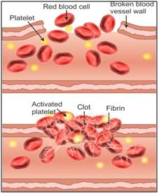
Solution D.7
(a) Pulmonary semilunar valves: They are located at the opening of the right ventricle into the pulmonary artery.
(b) Tonsils: They are located on the sides of the neck.
(c) Heart: It is located in the centre of the body between the two lungs and above the diaphragm.
(d) Pacemaker: It is present near the opening of the superior vena cava.
(e) Hepatic portal vein: It is located between the small intestine and the liver, in the upper right quadrant of the abdomen, which originates behind the neck of the pancreas.
Solution E.1
(a) 1 → Red Blood Cell (RBC),
2 → White Blood Cell (WBC),
3 → Blood Platelet
4 → Blood Plasma.
(b) The red blood cells (1) are minute biconcave disc-like structures whereas the white blood cells (2) are amoeboid.
(c) Function of part 1 (RBC): Transport of respiratory gases to the tissues and from the tissues, transport of nutrients from the alimentary canal to the tissues.
Function of part 2 (WBC): WBCs play major role in defense mechanism and immunity of the body.
Function of part 3 (Blood Platelet): Blood platelets are the initiator of blood clotting.
(d) The average life span of a red blood cell, RBC (1) is about 120 days.
(e) Thromboplastin
Solution E.2
(a) The structure 3 represents the heart. It forms the centre of double circulation and is located between the liver and the head (as per the diagram). Also the blood circulation (indicated by 1) begins from heart to lungs.
(b)
| Aorta | 5 |
| Hepatic portal vein | 7 |
| Pulmonary artery | 1 |
| Superior vena cava | 9 |
| Renal vein | 8 |
| Stomach | 10 |
Solution E.3
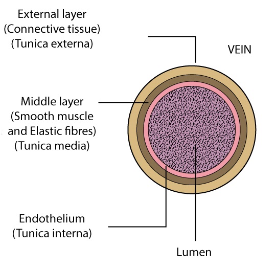
(a) A- Artery, B-Vein, C-Capillary
(b) 1 - External layer made of connective tissue
2 - Lumen
3 - Middle layer of smooth muscles and elastic fibres
4 - Endothelium
(c) An artery has thick muscular walls and a narrow lumen. It does not have any valve. A vein on the other hand has thin muscular walls and a wider lumen. It has valves to prevent backflow of blood.
(d) A (Artery)- Oxygenated blood, B (Vein)- Deoxygenated blood
(e) At the capillary level the actual exchange of gases takes place.
Solution E.4
(a) Atrial Diastole and Ventricular Systole
(b) Ventricular muscles are contracting during this phase because the valves between the two ventricles and pulmonary artery and aorta are open while the atrio-ventricular valves are closed.
(c)
|
1 |
Pulmonary Artery |
|
2 |
Aorta |
|
3 |
Pulmonary Vein |
|
4 |
Left Atrium |
|
5 |
Bicuspid Valve |
|
6 |
Right Ventricle |
(d) Part 1 (Pulmonary artery) → Deoxygenated blood
Part 2 (Aorta) → Oxygenated Blood
(e) Two i.e., bicuspid and tricuspid valves are closed in this phase.
Solution E.5
a. 1 - Red blood cell
b. Diapedesis
c.
|
RBC |
WBC |
|
They lack a nucleus. |
They have a nucleus. |
|
They are biconcave and disc-shaped. |
They are spherical and have different sizes. |
d. The process which occurs in B and C is phagocytosis. In this process, the WBCs engulf the foreign particles and destroy them, thus preventing the occurrence of disease.
Solution E.6
(a) 1- Right atrium, 2- Left ventricle, 3- Aorta, 4- Body parts (Whole body)
(b) Aorta (3) is the thickest artery.
(c) Left ventricle (2) has the thickest muscular wall.
(d) Whole body (body parts) (4) has the maximum number of blood capillaries.
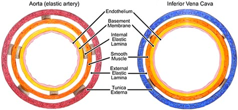
(e) Transverse section of vena cava and aorta (3):
Solution E.7
(a) The kind of blood vessels shown in the figure are veins. The branches are termed as venules.
(b) The structure shown inside the blood vessel is a valve. Valves prevent the backflow of the blood.
(c) Normally, deoxygenated blood flows through the veins. Arteries carry blood from the heart to the lungs.
(d) Hepatic vein and renal vein respectively.
(e) Transverse section of a vein:
Solution E.8
(a) 1- Aorta, 2- Left atrium, 3- Left ventricle, 4- Dorsal aorta, 5- Inferior vena cava, 6- Superior vena cava
(b) Inferior vena cava (5) brings blood from the posterior or the lower region of the body including abdomen and legs. Superior vena cava (6) brings deoxygenated blood from the anterior or upper regions of the body including head, chest, and arms.
(c) Left atrium (2) collects blood from the lungs through the blood vessel. The blood vessel is pulmonary vein.
(d) Differences between dorsal aorta and inferior venacava:
|
Dorsal aorta |
Inferior venacava |
|
• It has no valves and a narrow lumen. |
• It contains valves and a wider lumen. |
|
• It carries oxygenated blood and transports it to the lower part of the body tissues. |
• It collects deoxygenated blood from the lower part of the body and pours it into the right atrium. |
(e) When there is a blockage in any coronary artery or in any one or more of their branches, there is a deadening of the corresponding area of the heart muscles leading to myocardial infarction or heart attack.