Class 11-science NCERT Solutions Biology Chapter 7 - Structural Organisation In Animals
84
Structural Organisation In Animals Exercise 84
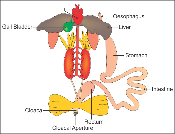
Solution 1
Digestive system of frog:
Solution 2
Function of Ureters in Frog:
In male frog, the ureters conduct both urine and spermatozoa to the cloaca. Hence, it acts as a urinogenital duct in male frogs. In females, the ureters conduct only urine and the oviducts open separately in the cloaca.