Class 11-science NCERT Solutions Biology Chapter 18 - Neural Control And Coordination
Neural Control And Coordination Exercise 237
Solution 1
Structure of the brain:
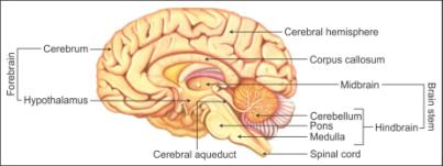
- The human brain is protected in the skull.
- The brain is covered by three membranes called cranial meninges.
- The outer layer of the meninges is called dura mater. It is a tough and fibrous membrane.
- The middle layer is called arachnoid. It is thin and delicate.
- The inner layer is the pia mater. It is in continuation with the brain tissue. It is highly vascular and richly supplied with blood.
- Three primary regions of the brain are
- Forebrain
- Midbrain
- Hindbrain
- Forebrain:
- The forebrain consists of cerebrum, thalamus and hypothalamus.
- Cerebrum:
- It forms the major part of the brain.
- The cerebrum is divided into halves longitudinally by a deep cleft. Each half is called the cerebral hemisphere.
- Both hemispheres are connected by the corpus callosum. It is a tract of nerve fibres.
- Cerebral hemispheres are hollow internally.
- The walls of the cerebrum have an outer cortex and inner medulla.
- The cerebral cortex contains cell bodies of neurons and hence appears greyish. It is called grey matter.
- The grey matter is thrown into many grooves and folds called sulci and gyri, respectively.
- A higher number of convolutions leads to greater intelligence.
- The cerebral cortex contains motor areas, sensory areas and association areas. Association areas are neither sensory nor motor. These areas are responsible for complex functions such as memory, communication and intersensory associations.
- The cerebral medulla consists of axons of nerve fibres and appears whitish. It is called white matter.
- The inner part of the cerebral hemispheres and a group of associated deep structures such as hippocampus and amygdala form a complex structure called the limbic lobe or limbic system.
- Functions:
- The cerebrum is the centre of intelligence, memory, consciousness, will power and voluntary actions.
- Thalamus:
- It is made of grey matter.
- It is situated superior to the midbrain.
- Functions:
- The thalamus relays motor and sensory impulses to the cerebrum.
- It also regulates the manifestation of emotions and recognises heat, cold and pain.
- Hypothalamus:
- It lies at the base of the thalamus.
- It consists of optic chiasma, a point where the fibres of optic nerves cross to opposite sides.
- Behind the optic chiasma is the infundibulum. It is a greyish protuberance of the hypothalamus.
- The infundibulum holds the pituitary gland.
- Functions:
- Hypothalamus contains the centres which control body temperature, blood pressure and homeostasis.
- It contains the centres to control hunger, thirst, sleep, fatigue, emotions, anger, pleasure and penance.
- The neurosecretory cells of the hypothalamus secrete certain hormones or releasing factors which control the activity of the pituitary hormones.
- The hypothalamus along with the limbic system is involved in the regulation of sexual behaviour.
- Midbrain:
- The midbrain consists of cerebral peduncles and corpora quadrigemina.
- Cerebral Peduncles:
- Cerebral peduncles are fibrous thick tracts.
- They connect the cerebrum and the cerebellum.
- Functions:
- They relay the sensory and motor impulse between the forebrain and hindbrain.
- Corpora Quadrigemina:
- Two pairs of solid lobes are present on the dorsal portion of the brain. These lobes are called corpora quadrigemina.
- One pair is called superior colliculi and the other pair is called inferior colliculi.
- Functions:
- The corpora quadrigemina controls the visual reflexes. They control the movement of the head and eye.
- They also control the auditory reflexes. They control the movement of the head to locate and detect the source of sound.
- Hindbrain:
- The hindbrain consists of the cerebellum, pons varolii and medulla oblongata.
- Cerebellum:
- It is located at the base of the cerebellum.
- The outer cerebellar cortex is made of grey matter and the inner cerebellar medulla is made of white matter.
- The white matter has fibre tracts which connect the cerebellum with the medulla oblongata and the cerebrum.
- Functions:
- It coordinates the muscular activity and the balance of the body.
- The impulse of performing muscular activity originates in the cerebrum.
- It modulates the voluntary movements initiated in the cerebrum.
- Pons varolii:
- It is made of a thick bundle of white nerve fibres.
- It lies above the medulla oblongata.
- Functions:
- It coordinates between the two lobes of the cerebellum.
- The pneumotaxic centre which controls breathing is located in the pons varolii.
- Medulla oblongata:
- It is located at the base of the skull.
- It is conical in shape.
- It continues behind the brain as the spinal cord.
- Injury to the medulla oblongata results in death.
- Functions:
- It acts as a pathway and conducts impulses from the spinal cord to the brain.
- It controls the activities of the internal organs, heartbeat and breathing.
Solution 2(a)
|
Central Neural System (CNS) |
Peripheral Neural System (PNS) |
| 1. CNS is made of the brain and the spinal cord. | 1. PNS consists of cranial nerves and spinal nerves. |
| 2. Brain is protected by the skull and the spinal cord by the vertebral column. | 2. There are no protective structures for PNS. |
| 3. CNS does not have any subdivisions. | 3. PNS is divided into somatic nervous system and autonomic nervous system. |
| 4. CNS processes the information and controls the response to impulses. | 4. Nerves of PNS carry impulses to the CNS and responses from the CNS to different organs of the body. |
| 5. Group of neurons in CNS is called nuclei. | 5. Group of neurons in PNS is called ganglia. |
Solution 2(b)
|
Resting Potential |
Action Potential |
|
1. It is the potential difference (difference in electric charge) across the membrane when the neuron is at the resting phase. |
1. It is the potential difference (difference in the electric charge) across the membrane when the neuron is stimulated or excited. |
|
2. The outer side of the neuron is positively charged and the interior of the neuron is negatively charged. |
2. The outer side of the neuron is negatively charged and the interior of the neuron is positively charged. |
|
3. The plasma membrane of the neuron is more permeable to K+ ions. |
3. The plasma membrane of the neuron is more permeable to Na+ ions. |
|
4. The sodium-potassium-ATPase pump is active and sends Na+ ions outside the neuron to maintain the resting potential. |
4. The sodium-potassium-ATPase pump functions in a reverse manner and sends Na+ ions into the neuron. |
Solution 3(a)
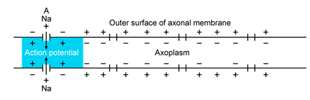
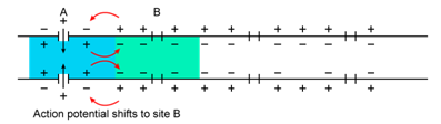
Polarisation of the membrane of a nerve fibre:
- When the nerve fibre is at the resting phase, it is said to be in the polarised state.
- In a polarised state, the membrane of the nerve fibre experiences resting potential.
- The following steps take place during the process of polarisation of the membrane of a nerve fibre:
- When a depolarised region of a nerve fibre starts becoming polarised initially, there are more K+ ions outside the nerve fibre and the axon membrane contains large amount of Na+ ions.
- As the region of the membrane starts attaining the polarised state, the membrane becomes more permeable to K+ ions and impermeable to Na+ ions and negatively charged proteins.
- 3 Na+ ions are sent outside the axon and 2 K+ ions are sent into the axon by a sodium-potassium pump by active transport.
- The inner side of the membrane becomes electronegative (negatively charged) and the outer side becomes electropositive (positively charged) because of the movement of sodium and potassium ions. This makes the nerve fibre polarised.
Solution 3(b)
Depolarisation of the membrane of a nerve fibre:
- When the nerve fibre is stimulated, it is said to be in the depolarised state.
- In a depolarised state, the membrane of the nerve fibre experiences an action potential.
- The following steps take place during the process of depolarisation of the membrane of a nerve fibre:
- In a polarised state, the axon has more concentration of K+ ions and outside the axon, the concentration of Na+ ions is more.
- When the nerve fibre gets excited by the stimulus, the permeability of the membrane for Na+ ions and K+ ions is reversed.
- The membrane becomes highly permeable for Na+ ions.
- There is a rapid influx of Na+ ions into the axon.
- This make the inner side of the membrane positively charged and the outside of the membrane becomes negatively charged.
- This results in depolarisation of the membrane of the nerve fibre and its experiences an action potential.
Solution 3(c)
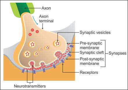
Transmission of a nerve impulse across a chemical synapse:
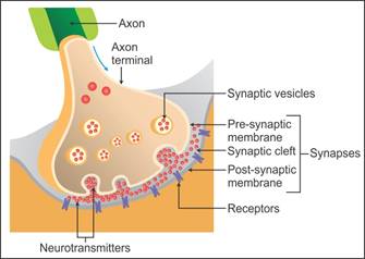
- A synapse is formed by the membranes of the pre-synaptic neuron and the post-synaptic neuron.
- A synapse may or may not be separated by a gap which is called the synaptic cleft.
- At a chemical synapse, the pre-synaptic and post-synaptic neurons are separated by the synaptic cleft.
- When an impulse arrives at the axon terminal, the calcium ions present in the synaptic cleft enter the synaptic knobs present at the axon terminals of the pre-synaptic neuron.
- The synaptic vesicles in the synaptic knobs of the pre-synaptic neuron move towards the plasma membrane and fuse with it.
- The vesicles release the neurotransmitter acetylcholine in the synaptic cleft. (Empty synaptic vesicles return to the cytoplasm of the pre-synaptic neuron where they are refilled.)
- The molecules of the acetylcholine bind to the protein receptors present on the plasma membrane of the post-synaptic neurons.
- This binding opens the channels and sodium ions enter the post-synaptic neuron, while potassium ions leave the post-synaptic membrane.
- This generates an action potential in the membrane of the post-synaptic neuron, and hence, the impulse is transmitted to the post-synaptic neuron.
Neural Control And Coordination Exercise 238
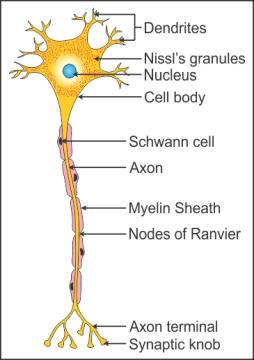
Solution 4(a)
Neuron:
Solution 4(b)
Brain:

Solution 5(a)
Neural coordination:
- Coordination is a characteristic feature of living organisms.
- Coordination is the process through which two or more organs interact and complement the functions of one another.
- Coordination is achieved by two ways in humans and other higher order animals—neural coordination and chemical coordination.
- Neural coordination is carried out by highly specialised cells called neurons.
- The neural system is a network of point-to-point connections between the neurons and the organs and it operates through nerve impulses.
- Neural coordination is always between the stimulus and the response—receptors and effectors.
- All body functions are carried out and controlled by neural coordination.
- The stimulus is received from organs such as the skin and a response is generated which is sent to the muscles or glands.
- The previous stimulus is always stored in memory by the neural system.
- Neural coordination helps in controlling and harmonising voluntary actions such as running, walking, writing and talking.
- It helps us to remember, analyse, think and reason because the brain, a part of the neural system, is the site of intelligence.
- All vital functions such as breathing, working of the heart and digestion are controlled by neural coordination.
- It helps maintain homeostasis by coordinating between various metabolic activities of the body.
Solution 5(b)
Forebrain:
- The forebrain consists of cerebrum, thalamus and hypothalamus.
- Cerebrum:
- The forebrain
- It forms the major part of the brain.
- The cerebrum is divided into halves longitudinally by a deep cleft. Each half is called a cerebral hemisphere.
- Both hemispheres are connected by the corpus callosum-a tract of nerve fibres.
- The cerebral hemispheres are hollow internally.
- The walls of the cerebrum have an outer cortex and an inner medulla.
- The cerebral cortex contains cell bodies of neurons and hence appears greyish. It is called grey matter.
- The grey matter is thrown into many grooves and folds called sulci and gyri, respectively.
- A higher number of convolutions leads to greater intelligence.
- The cerebral cortex contains motor areas, sensory areas and association areas. Association areas are neither sensory nor motor. These areas are responsible for complex functions such as memory, communication and intersensory associations.
- The cerebral medulla consists of axons of nerve fibres and appears whitish. It is called white matter.
- The inner part of the cerebral hemispheres and a group of associated deep structures such as hippocampus and amygdala form a complex structure called the limbic lobe or limbic system.
- Functions:
- The cerebrum is the centre of intelligence, memory, consciousness, will power and voluntary actions.
- Thalamus:
- It is made of grey matter.
- It is situated superior to the midbrain.
- Functions:
- The thalamus relays motor and sensory impulses to the cerebrum.
- It also regulates the manifestation of emotions and recognises heat, cold and pain.
- Hypothalamus:
- It lies at the base of the thalamus.
- It consists of the optic chiasma, a point where the fibres of optic nerves cross to opposite sides.
- Behind the optic chiasma is the infundibulum. It is a greyish protuberance of the hypothalamus.
- The infundibulum holds the pituitary gland.
- Functions:
- The hypothalamus contains the centres which control body temperature, blood pressure and homeostasis.
- It contains the centres to control hunger, thirst, sleep, fatigue, emotions, anger, pleasure and penance.
- The neurosecretory cells of the hypothalamus secrete certain hormone or releasing factors which control the activity of the pituitary hormones.
- The hypothalamus along with the limbic system is involved in the regulation of sexual behaviour.
Solution 5(c)
Midbrain:
- The midbrain consists of cerebral peduncles and corpora quadrigemina.
- Cerebral Peduncles:
- Cerebral peduncles are fibrous thick tracts.
- They connect the cerebrum and the cerebellum.
- Functions:
- They relay the sensory and motor impulse between the forebrain and hindbrain.
- Corpora Quadrigemina:
- On the dorsal portion of the brain, there are two pairs of solid lobes present. These lobes are called corpora quadrigemina.
- One pair is called superior colliculi and the other pair is called inferior colliculi.
- Functions:
- The corpora quadrigemina controls the visual reflexes. They control the movement of the head and the eye.
- They also control auditory reflexes. They control the movement of the head to locate and detect the source of sound.
Solution 5(d)
Hindbrain:
- The hindbrain consists the cerebellum, pons varolii and medulla oblongata.
- Cerebellum:
- It is located at the base of the cerebellum.
- The outer cerebellar cortex is made of grey matter and the inner cerebellar medulla is made of white matter.
- The white matter has fibre tracts which connect the cerebellum with the medulla oblongata and the cerebrum.
- Functions:
- It coordinates muscular activity and the balance of the body.
- The impulse of performing muscular activity originates in the cerebrum.
- It modulates the voluntary movements initiated in the cerebrum.
- Pons varolii:
- It is made of a thick bundle of white nerve fibres.
- It lies above the medulla oblongata.
- Functions:
- It coordinates the two lobes of the cerebellum.
- The pneumotaxic centre which controls breathing is located in the pons varolii.
- Medulla oblongata:
- It is located at the base of the skull.
- It is conical in shape.
- It continues behind the brain as the spinal cord.
- Injury to the medulla oblongata results in death.
- Functions:
- It acts as a pathway and conducts impulses from the spinal cord to the brain.
- It controls the activities of the internal organs, heartbeat and breathing.
Solution 5(e)
Synapse:
- A synapse is formed by the membranes of the pre-synaptic neuron and the post-synaptic neuron.
- A synapse may or may not be separated by a gap which is called the synaptic cleft.
- There are two kinds of synapses-electrical synapse and chemical synapse.
- Mechanism of electrical synapse:
- In this case, the pre-synaptic and post-synaptic membranes are in proximity.
- Impulse in the form of electric current directly flows from the pre-synaptic neuron to the post-synaptic neuron.
- Transmission is faster than the chemical synapse.
- Mechanism of chemical synapse:
- Here, the pre-synaptic and post-synaptic neurons are separated by the synaptic cleft.
- When an impulse arrives at the axon terminal, the calcium ions present in the synaptic cleft enter the synaptic knobs present at the axon terminals of the pre-synaptic neuron.
- The synaptic vesicles in the synaptic knobs of the pre-synaptic neuron move towards the plasma membrane and fuse with it.
- The vesicles release the neurotransmitter acetylcholine in the synaptic cleft. (Empty synaptic vesicles return to the cytoplasm of the pre-synaptic neuron where they are refilled.)
- The molecules of acetylcholine bind to the protein receptors present on the plasma membrane of the post-synaptic neurons.
- This binding opens the channels, and sodium ions enter the post-synaptic neuron, while potassium ions leave the post-synaptic membrane.
- This generates an action potential in the membrane of the post-synaptic neuron, and hence, the impulse is transmitted to the post-synaptic neuron.
Solution 6
Mechanism of synaptic transmission:
- A synapse is formed by the membranes of the pre-synaptic neuron and the post-synaptic neuron.
- A synapse may or may not be separated by a gap called the synaptic cleft.
- There are two kinds of synapses—electrical synapse and chemical synapse.
- Mechanism of synaptic transmission at the electrical synapse:
- In this case, the pre-synaptic and post-synaptic membranes are in proximity.
- Impulse in the form of electric current directly flows from the pre-synaptic neuron to the post-synaptic neuron.
- Transmission is faster than the chemical synapse.
- Mechanism of synaptic transmission at the chemical synapse:
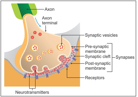
- Here, the pre-synaptic and post-synaptic neurons are separated by the synaptic cleft.
- When an impulse arrives at the axon terminal, the calcium ions present in the synaptic cleft enter the synaptic knobs present at the axon terminals of the pre-synaptic neuron.
- The synaptic vesicles present in the synaptic knobs present in the pre-synaptic neuron move towards the plasma membrane and fuse with it.
- The vesicles release the neurotransmitter acetylcholine in the synaptic cleft. (Empty synaptic vesicles return to the cytoplasm of the pre-synaptic neuron where they are refilled.)
- The molecules of the acetylcholine bind to the protein receptors present on the plasma membrane of the post-synaptic neurons.
- This binding opens the channels, and sodium ions enter the post-synaptic neuron, while potassium ions leave the post-synaptic membrane.
- This generates an action potential in the membrane of the post-synaptic neuron, and hence, the impulse is transmitted to the post-synaptic neuron.
Solution 7
In a stimulated nerve fibre, sodium channels of the neurilemma get activated and open. Sodium ions diffuse from the outside to the intracellular fluid because of the electrochemical gradient. The potassium ions move out, and the membrane becomes negatively charged from outside and positively charged from inside. This sudden change in the membrane potential is called the action potential, and the membrane is said to be depolarised.
Solution 8(a)
|
Myelinated Axon |
Non-myelinated Axon |
| 1. Myelin sheath is present. | 1. Myelin sheath is absent. |
| 2. Nodes of Ranvier are present. | 2. Nodes of Ranvier are absent. |
| It is found in the grey matter of the brain, spinal cord and autonomous nervous system. | 3. It is found in the white matter of the brain, spinal cord and autonomous nervous system. |
| 4. The conduction of nerve impulse is node to node. | 4. The conduction of nerve impulse is smooth. |
| 5. The speed of conduction of impulse is 50 times faster than in non-myelinated axon. | 5. The speed of conduction of impulse is slow. |
Solution 8(b)
|
Dendrites |
Axons |
|
1. They are short processes. |
1. Axons are long processes. |
|
2. Dendrites carry impulses towards the cell body of the neuron. |
2. Axons carry impulses away from the cell body of the neuron. |
|
3. Dendrites are always branched. |
3. Axons may or may not be branched. |
|
4. Nissl's granules are present in the neuroplasm. |
4. Nissl's granules are absent in the neuroplasm. |
Solution 8(c)
|
Thalamus |
Hypothalamus |
|
1. It is made of only grey matter. |
1. It is made of white and grey matter. |
|
2. The thalamus does not secrete any hormone. |
2. The hypothalamus secretes certain hormones which control the activity of the pituitary gland. |
|
3. It is situated superior to the midbrain. |
3. It is situated at the base of the thalamus. |
|
4. It contains the centres of sensations such as heat, cold and pain. |
4. It contains the centres which control body temperature, blood pressure and homeostasis. |
Solution 8(d)
|
Cerebrum |
Cerebellum |
|
1. It is the largest part of the brain. |
1. It is the second largest part of the brain. |
|
2. It is part of the forebrain. |
2. It is part of the hindbrain. |
|
3. The cerebrum is divided into two cerebral hemispheres. |
3. The cerebellum is divided into three lobes-central vermis and the two lateral cerebellar hemispheres. |
|
4. It is the site of memory and intelligence. |
4. It is the site of body equilibrium and posture. |
Solution 9
(a) The cerebrum is the most developed.
(b) The hypothalamus of the central neural system acts as a master clock.
Solution 10(a)
|
Afferent Neurons |
Efferent Neurons |
| 1. They conduct sensory impulses from the receptors to the central nervous system. | 1. They conduct motor impulses from the central nervous system to the effector organs such as muscles. |
| 2. They are present in the sense organs. | 2. They are present in the brain and the spinal cord. |
| 3. They are sensory neurons. | 3. They are motor neurons. |
Solution 10(b)
|
Impulse Conduction in a Myelinated Nerve Fibre |
Impulse Conduction in Non-myelinated Nerve Fibre |
|
1. Impulse travels from node to node. |
1. Impulse travels along the length of the entire nerve fibre. |
|
2. The speed of conduction is 50 times faster than the non-myelintaed nerve fibre. |
2. The speed of conduction is slower. |
|
3. Energy expenditure during impulse transmission is less. |
3. Energy expenditure during impulse transmission is more. |
Solution 10 (c)
|
Cranial Nerves |
Spinal Nerves |
|
1. There are 12 pairs of cranial nerves. |
1. There are 31 pairs of spinal nerves. |
|
2. They arise from the brain and extend to the other parts of the body. |
2. They arise from the spinal cord and extend to other parts of the body. |
|
3. They may be sensory, motor or mixed. |
3. They are mixed nerves. |