Class 10 SELINA Solutions Biology Chapter 11 - Sense Organs
Sense Organs Exercise Ex. 1
Solution A.1
(d) Retina
Solution A.2
(a) Vitamin A
Solution A.3
(d) Iris
Solution A.4
(c) Lysozyme
Solution A.5
(a) Iris
Solution A.6
(c) Endolymph
Solution A.7
(a) Cornea
Solution A.8
(c) Cochlea
Solution A.9
(d) Organ of Corti
Solution A.10
(b) Eustachian tube
Solution B.1
(a) Rhodopsin
(b) Eustachian tube
(c) Hammer
(d) Eustachian tube
(e) Cornea
(f) Auditory nerves
(g) Rods and cones
(h) Hypermetropia
Solution B.2
(a) Cones : Iodopsin :: Rods : Rhodopsin
(b) Eyes : Photoreceptors :: Ears : Phonoreceptors
(c) Ears : Auditory nerve :: Eyes : Optic nerve
(d) Ear pinna : Auricle :: Inner ear : Membranous labyrinth
(e) Semi-circular canal : Ampulla :: Cochlea : Organ of Corti
Solution B.3
|
Column I |
Column II |
|
(i) Conjunctiva |
(d) Transparent epithelium |
|
(ii) Cornea |
(g)Transparent but appears black |
|
(iii) Choroid |
(f) Contains melanin |
|
(iv) Cochlea |
(c) Spiral shaped |
|
(v) Conjunctivitis |
(a) Viral infection |
Solution C.1
(a) False
Correct statement: Deafness is caused due to rupturing of the eardrum.
(b) False
Correct statement: Semicircular canals are concerned with dynamic balance.
Solution C.2
(a) Yellow spot: It lies at the back of the eye almost at the centre on the horizontal axis of the eyeball.
(b) Lacrimal gland: It is located at the upper sideward portion of the eye orbit.
(c) Organ of Corti: It is located within the cochlea between the vestibular duct and the tympanic duct.
(d) Eustachian canal: It connects the anterior wall of the middle ear to the lateral wall of the throat.
(e) Incus: It is located in the middle ear between the malleus and the stapes.
Solution C.3
(a) Auditory canal, tympanum, ear ossicles, oval window, cochlea
(b) Conjunctiva, cornea, lens, retina, optic nerve
Solution C.4
(a) Cochlea: It contains Organ of Corti which contains sensory cells for hearing. They transmit sound impulses to the brain through the auditory nerve.
(b) Semicircular canals: They contain sensory cells for dynamic balance while the body is in motion and the nerve fibres from these canals join the auditory nerve.
(c) Iris: It contains radial muscles to widen and circular muscles to constrict the pupil, thereby regulating the amount of light entering the eye.
(d) Choroid: It is richly supplied with blood vessels for providing nourishment to the eye.
(e) Ciliary body and suspensory ligament: The ciliary body contains smooth muscles, which on contraction and relaxation, change the shape of the lens for viewing objects at different locations.
Solution C.5
|
Structure |
Function |
|
Auditory nerve |
Transfers impulse from inner ear to brain |
|
Ciliary muscle |
Helps to change the focal length of the eye lens |
|
Semicircular canals |
Dynamic equilibrium |
Solution C.6
(a) Rhodopsin and iodopsin.
(b) Dark adaptation and light adaptation.
(c) Distant vision accommodation and near vision accommodation.
(d) Sclera, choroid and retina.
Solution C.7
|
Cause |
Eye defect |
|
(a) Lens turns opaque |
Cataract |
|
(b) Uneven curvature of the cornea |
Astigmatism |
|
(c) Deficiency of vitamin A |
Night blindness |
|
(d) Lens loses its flexibility |
Presbyopia |
|
(e) Eye ball lengthens from front to back |
Myopia |
|
(f) Lens becomes too flat |
Hyperopia |
Solution D.1
(a) Conjunctiva: Conjunctiva is a thin membrane covering the entire front part of the eye and is continuous with the inner lining of the eyelids.
(b) Lysozyme: Lysozyme is a special enzyme found in the tears of the eyes. It has an antiseptic property which kills the germs.
(c) Adaptation: Adaptation is the ability of the eye to adjust to varying levels of darkness and light.
(d) Power of accommodation: The process of focusing the eye to see objects at different distances is called power of accommodation.
(e) Ear ossicles: The three tiny bones in the middle - the malleus, the incus, and the stapes are collectively referred to as the ear ossicles.
Solution D.2
(a) Differences between myopia and hyperopia (type of lens used for correction):
|
Myopia |
Hyperopia |
|
Myopia can be corrected by suitable concave (diverging) lens. |
Hyperopia can be corrected by suitable convex (converging) lens. |
(b) Differences between rods and cones (sensitivity):
|
Rods |
Cones |
|
Rods are sensitive to dim light but do not respond to colour. |
Cones are sensitive to bright light and are responsible for colour vision. |
(c) Differences between aqueous humour and vitreous humour (location):
|
Aqueous humour |
Vitreous humour |
|
Aqueous humour is the front chamber between the lens and the cornea. |
Vitreous humour is larger cavity of the eyeball behind the lens. |
(d) Differences between near and distant accommodation (shape of lens):
|
Near accommodation |
Distant accommodation |
|
For near accommodation, the lens becomes more convex or rounded. |
For distant accommodation, the lens is more flattened or thinner. |
(e) Differences between dark and light adaptation (pigments which will be regenerated):
|
Dark adaptation |
Light adaptation |
|
For dark adaptation, visual purple or rhodopsin pigment will be regenerated. |
For light adaptation, visual violet or iodopsin pigment will be regenerated. |
(f) Differences between night blindness and colour blindness (sensory cells which cannot function properly):
|
Night blindness |
Colour blindness |
|
In night blindness, the rod cells cannot function properly. |
In colour blindness, the cone cells cannot function properly. |
Solution D.3
(a) Sometimes medicines dropped into the eyes come into the nose and even throat because the nasolacrimal duct conducts the secretion into the nasal cavity.
(b) Three small bones of ear ossicles transmit the vibrations received by the tympanum and amplify them. If these were replaced by a single bone, the vibrations received by the tympanum would not be amplified. Hence, three small bones of ear ossicles are advantageous as compared to one single bone for hearing.
(c) There are no sensory cells in the blind spot and therefore, this is considered as 'area of no vision' and image striking it cannot be perceived.
Solution D.4
The image formed on the retina is real and inverted.
Solution D.5
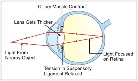
While reading a book, the lens is more convex or rounded due to contraction of ciliary muscles because the book is usually read from a short distance. When we raise our head and look at a distant object, the ciliary muscles relax to build the tension on the suspensory ligament so that they can stretch the lens. This change in the curvature of the lens makes us focus on distant object.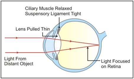
Solution D.6
If we look at a bright object and then close our eyes, the sensation of light persists for a short period. This is known as persistence image or the after image. It lasts for one-tenth of a second. Therefore by closing the eyes and gently pressing them with your palms, you see some specs of brilliant light.
Solution D.7
The three ear ossicles are: Malleus (hammer), Incus (anvil) and Stapes (stirr up).
The last ear ossicle, stapes, vibrates and transmits the vibration to the oval window.
The role of other two ear ossicles is to magnify the vibration of stapes as a result of their lever like action.
Solution E.1
(a) The adjustment done in figure A is dark adaptation whereas in figure B, it is light adaptation.
(b) Differences between the adjustments in figures (A) and (B):
|
On the basis of |
Dark adaptation (A) |
Light adaptation (B) |
|
Size of the pupil |
Dilated |
Constricted |
|
Pigment which gets generated |
Rhodopsin |
Iodopsin |
|
Cells of the retina |
Cones become inactive and disintegration of iodopsin starts |
Rods become inactive and disintegration of rhodopsin starts |
Solution E.2
(a)
(i) Iris
(ii) Ciliary body
(iii) Aqueous humour
(iv) Choroid layer
(b)
(i) Utriculus and sacculus
(ii) Semi-circular canals
(iii) Cochlea
(iv) Ear ossicles
Solution E.3
(a) 1 - Aqueous chamber, 2 - Lens, 3 - Iris, 4 - Cornea, 5 - Conjunctiva, 6 - Sclera, 7 - Choroid, 8 - Retina, 9 - Yellow spot, 10 - Optic nerve (Blind spot)
(b) Part 3 (Iris) - It contains radial muscles to dilate the pupil and circular muscles to constrict the pupil.
Part 7 (Choroid) - It is the middle layer of the eyeball, richly supplied with blood vessels and provides nourishment to the eye.
(c) Part 9 (yellow spot) contains sensory cells especially the cone cells while part 10 (blind sport) contains no sensory cells.
(d) Part 6 (sclera) gives shape to the eyeball and part 8 (retina) acts as screen to form image of an object.
Solution E.4
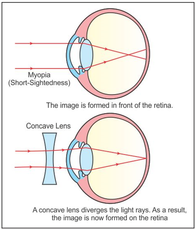
(a) Myopia
(b) Possible reasons for myopia:
- The eye ball is lengthened from front to back
- The lens is too curved
(c) 1 - vitreous humour, 2 - blind spot, 3-lens, 4-pupil
(d) Concave lens
(e) Myopia and its correction using a concave lens
Solution E.5
(a) Membranous labyrinth in the inner ear
 (b)
(b)
(i) Cochlea
(ii) Organ of Corti
(iii) Oval window
(iv) Auditory nerves
(v) Eustachian tube
Solution E.6
(i) Ear ossicles
(ii) A - Cochlea, B - Semicircular canals, C - Ear ossicles
(iii) Cochlea helps in transmitting impulses to the brain via the auditory nerve. Semicircular canals help in maintaining dynamic equilibrium of the body.
(iv) Organ of Corti
Solution E.7
Inner ear

Utriculus and Sacculus are responsible for maintaining static balance in human beings.
Solution E.8
(a) Myopia
(b) A-Normal eye, B-Myopia
(c) Looking glasses with the concave lens are required here.
Solution E.9
(a) 1 - External ear (pinna), 2 - Ear drum (tympanum), 3 - Auditory canal, 4 - Malleus, 5 - Semicircular canals, 6 - Cochlea, 7 - Auditory nerve, 8 - Eustachian tube.
(b) Part 6 (Cochlea) - It contains sensory cells for hearing.
Part 7 (Auditory nerve) - It transmits impulse of hearing to the brain.
Part 8 (Eustachian tube) - It equalizes air pressure on both the sides of the tympanum.
(c) It is harmful to use a sharp object to remove ear wax as it can rupture the ear drum.
The part involved is part 2 - Ear drum (tympanum).