Class 12-science NCERT Solutions Biology Chapter 2 - Human Reproduction
Human Reproduction Exercise 39
Solution 1
(a) Humans reproduce sexually.
(b) Humans are viviparous.
(c) Fertilisation is internal in humans.
(d) Male and female gametes are haploid.
(e) A zygote is diploid.
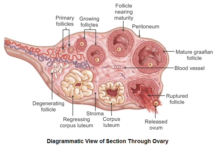
(f) The process of release of ovum from a mature follicle is called ovulation.
(g) Ovulation is induced by a hormone called luteinising hormone (LH).
(h) The fusion of male and female gametes is called fertilisation.
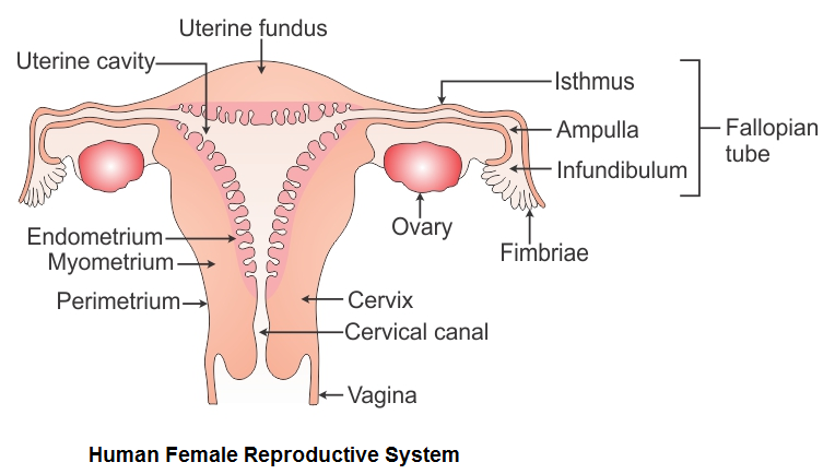
(i) Fertilisation takes place in ampulla of oviduct.
(j) The zygote divides to form blastocyst which is implanted in the uterus.
(k) The structure which provides a vascular connection between the foetus and the uterus is called placenta.
Human Reproduction Exercise 40
Solution 2
Solution 3
Solution 4
Functions of the testis:
- Production of sperms by the seminiferous tubules by the process of spermatogenesis.
- Secretion of the male sex hormone testosterone by Leydig cells.
Functions of the ovary:
- Production of ova by the ovaries by the process of oogenesis.
- Secretion of the female sex hormones oestrogen and progesterone.
Solution 5
Structure of the seminiferous tubule:
- Seminiferous tubules are highly coiled structures present in testicular lobules. Each testicular lobule consists of 1–3 highly coiled seminiferous tubules.
- The wall of each seminiferous tubule is lined with germinal epithelium.
- Majority of the cells of this epithelium are cubical and called male germ cells or spermatogonia and a few large cells are called Sertoli cells or nurse cells.
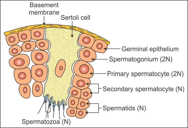
- The spermatogonia proliferate by mitosis and differentiate into primary spermatocytes, which undergo meiosis to produce secondary spermatocytes and finally spermatozoa.
- The regions outside the seminiferous tubules are called interstitial spaces. They are filled with connective tissue which includes blood vessels and Leydig cells.
Functions of Sertoli cells:
- Support the developing germ cells and provide nutrition to the spermatids.
- Secrete androgen-binding protein which concentrates testosterone in the seminiferous tubules.
- Secrete inhibin which suppresses FSH synthesis.
Functions of Leydig cells:
- They synthesise and secrete androgens or the male sex hormone testosterone.
Solution 6
Spermatogenesis is the process where diploid sperm mother cells or spermatogonia in the seminiferous tubules of the testes change into haploid, microscopic and motile male gametes or spermatozoa. It is a stepwise process and occurs continuously in the testes which lie outside the abdomen and in the scrotum of human males. Spermatogenesis includes the formation of spermatids and spermatozoa.
Formation of spermatids:
- Multiplicative phase: During this phase, the sperm mother cells are differentiated from the germinal epithelium of the seminiferous tubules of the testes. They divide repeatedly by several mitotic divisions to form several daughter cells. They are called spermatogonia.
- Growth phase: During this phase, the diploid spermatogonia undergo the process of spermatocytogenesis where they derive nourishment from the nursing cells. They grow and increase in size because of the accumulation of nutritive material. Each spermatogonium is called a primary spermatocyte which bears a diploid number of chromosomes. In this phase, the primary spermatocyte prepares itself for entering the maturation phase.
- Maturation phase: During this phase, the primary spermatocytes undergo two maturation divisions—the first meiotic division (meiosis I) differentiates the primary spermatocyte into two haploid secondary spermatocytes. The second meiosis (meiosis II) differentiates each secondary spermatocyte into two spermatids. Thus, four haploid spermatids are formed by each spermatocyte.
Formation of spermatozoa from spermatids:
- Later, spermatids are transformed into flagellated sperms by the process of spermiogenesis.
- Four sperms are formed from one single spermatogonium.
- After spermiogenesis, sperm heads become embedded in the Sertoli cells. They are released from the seminiferous tubules by spermiation.
Solution 7
Gonadotropin-releasing hormone (GnRH), luteinising hormone (LH), follicle-stimulating hormone (FSH), androgens and inhibin are some major hormones involved in the regulation of spermatogenesis.
- At the age of puberty, a hypothalamic hormone called gonadotropin-releasing hormone (GnRH) is secreted which acts at the anterior pituitary gland and stimulates the secretion of two gonadotropins—luteinising hormone (LH) and follicle-stimulating hormone (FSH).
- LH acts at the Leydig cells and stimulates synthesis and secretion of androgens.
- Androgens stimulate inhibin production which regulates the process of spermatogenesis.
- FSH acts on the Sertoli cells and stimulates the secretion of factors which help in the process of spermiogenesis.
Solution 8
Spermiogenesis is the process of transformation of non-motile spermatids into mature, motile spermatozoa.
Spermiation is the process of release of mature spermatozoa from the Sertoli cells into the lumen of the seminiferous tubules of the testes.
Solution 9
Solution 10
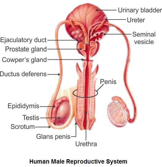
The seminal plasma or seminal fluid carries the secretions of the accessory sex glands of males, i.e. seminal vesicles, prostate gland and bulbourethral glands. Its main components are
- Fructose
- Calcium
- Certain enzymes
Solution 11
Male accessory ducts include the rete testis, vasa efferentia, epididymis and vasa deferentia.
|
Male accessory ducts |
Functions |
|
Rete testis |
|
|
Vasa efferentia |
|
|
Epididymis |
|
|
Vasa deferentia |
|
Male accessory glands include the seminal vesicles, prostate gland and bulbourethral glands.
|
Male accessory glands |
Functions |
|
Seminal vesicles |
|
|
Prostate gland |
|
|
Bulbourethral glands/Cowper’s glands |
|
Solution 12
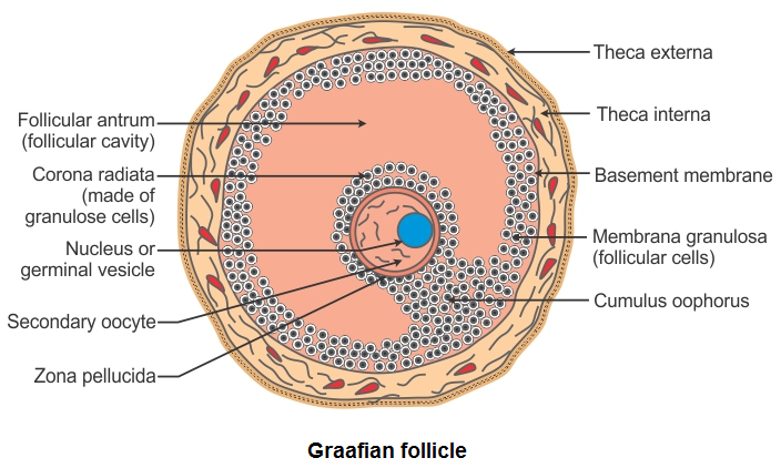
Oogenesis is the process of formation of haploid female gametes called ova from diploid oogonia in the Graafian follicles of the ovary. It is a discontinuous process which starts during the period of foetal development but is completed only after the onset of puberty.
Oogenesis occurs in the following three phases:
- Multiplicative phase: During this phase, the follicle cells are differentiated from the germinal epithelium of the ovary because of repeated mitosis division. Some follicle cells become large and are known as egg mother cells. They again divide by mitosis to increase their number. These are then called oogonia.
- Growth phase: During this phase, one of the oogonium of the egg nest differentiates and the remaining oogonia change into surrounding nutritive follicular epithelium. The differentiated isolated oogonium increases in size by obtaining nourishment from the surrounding follicle cells and transforms into a diploid primary oocyte.
- Maturation phase: During this phase, the diploid primary oocyte undergoes two maturation divisions. The first meiotic division (meiosis I) divides the diploid primary oocyte into two haploid cells. The larger one is the secondary oocyte and the smaller one is the polar body or polocyte. The secondary oocyte undergoes a second meiotic division (meiosis II) and forms one large ootid and a small second polar body. The first polar body also divides again by mitosis and forms two polar bodies. The ootid grows into a functional haploid ovum. Thus, from one primary oocyte, one large ovum and three polar bodies are formed. The three polar bodies degenerate as they do not take part in reproduction, and only one functional ovum is left behind.
Solution 13
Solution 14
Solution 15
(a) Corpus luteum: It acts as an endocrine gland and secretes the hormone progesterone which prepares the uterine endometrium for implantation, placentation and normal development of the foetus and thereby maintains pregnancy.
(b) Endometrium: It is the innermost, highly vascular and glandular lining of the uterus. It undergoes cyclic changes during the different phases of the menstrual cycle to prepare itself for the implantation of the blastocyst and placentation.
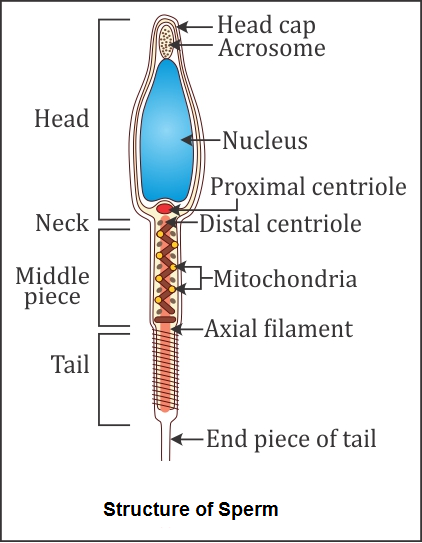
(c) Acrosome: It secretes sperm lysins such as hyaluronidase, corona penetrating enzyme and acrosin. These hydrolytic enzymes help contact the ovum, dissolve its membranes and finally penetrate it for fertilisation.
(d) Sperm tail: It helps in the movement of spermatozoon in the female genital tract for fertilisation. It produces lashing movements which provide forward push to the sperm.
(e) Fimbriae: These are finger-like projections present on the margins of the infundibulum. They bear cilia which beat towards the ostium for the collection of ovum from the ovary after ovulation.
Solution 16
(a) False. Androgens are secreted by Leydig cells or interstitial cells.
(b) True
(c) False. Leydig cells are found in regions called interstitial spaces outside the seminiferous tubules of the testis.
(d) True
(e) False. Oogenesis takes place in the Graafian follicle of the ovaries.
(f) True
(g) True
Solution 17
The menstrual cycle in human females starts at the age of puberty and carries on up to menopause. During this cycle, the ovaries, uterus and fallopian tubes undergo several regular cyclic changes because of the influence of the pituitary hormones so that the fertilised ovum is implanted. If fertilisation fails to occur, then the corpus luteum of the ovary degenerates and progesterone and oestrogen secretion stop, leading to a fresh start of the menstrual cycle. This cycle is usually repeated after 28 days and lasts for 3–5 days.
Role of hormones in the menstrual cycle:
|
Hormone |
Changes |
|
|
|
FSH |
|
|
|
|
LH |
|
|
|
|
Oestrogen |
|
|
|
|
Progesterone |
|
|
|
Solution 18
Parturition is the process of expulsion of the fully developed foetus from the mother’s uterus after the gestation period. Oxytocin and relaxin are involved in the induction of parturition.
Role of hormones in induction of parturition:
Oxytocin: It acts on the uterine muscles and induces forceful contractions of the myometrium of the uterus leading to the expulsion of the baby.
Relaxin: It causes relaxation of the pelvic ligaments and widens the pelvis to facilitate easy childbirth.
Solution 19
- Males are heterogametic, i.e. they produce two types of male gametes (sperms). 50% of sperms carry the X chromosome, while the other 50% carry the Y chromosome.
- Females are homogametic, i.e. they produce only one type of gamete (ova), each carrying the X chromosome.
- After fusion of the male and female gametes, the zygote would carry either XX or XY depending on whether the sperm carrying X or Y fertilised the ovum.
- The zygote carrying XX would develop into a female baby and XY would form a male baby.
- Thus, the sex of the baby is determined by the father and not by the mother.
- Therefore, it is not correct on the part of society to blame women for giving birth to daughters.
Solution 20
Generally, only one egg (rarely two) is released by human ovaries in a month.
In case of identical or monozygotic twins, one egg is released which divides into two after fertilisation, thus giving the same genetic features.
In case of fraternal or dizygotic twins, two eggs are released and get fertilised by two different sperms. So, the genetic characters of fraternal twins are always different.
Solution 21
To give birth to six puppies, six eggs must have been released by the ovary of a female dog. Therefore, six zygotes would have been formed, each of which developed into a puppy.