Class 11-science NCERT Solutions Biology Chapter 17 - Locomotion And Movement
Locomotion And Movement Exercise 228
Solution 1
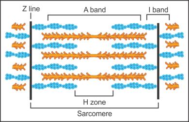
Different regions of a sarcomere of skeletal muscle:
Solution 2
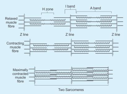
The theory which states that the contraction of a muscle fibre takes place because of the sliding of the thin filaments over the thick filaments is called the sliding filament theory.
Solution 3
Steps involved in muscle contraction:
1. Depolarisation of Sarcolemma:
- A signal sent by the central nervous system via a motor neuron reaches the neuromuscular junction or motor end plate.
- The neuromuscular junction is the junction between a motor neuron and the sarcolemma of the muscle fibre.
- The signal reaching the neuromuscular junction causes the release of acetylcholine, a neurotransmitter.
- Release of acetylcholine sets the action potential in the sarcolemma.
2. Release of Calcium Ions:
- Sarcolemma transmits its action potential to the sarcoplasmic reticulum to release calcium ions in the sarcoplasm.
3. Conformational Changes in Actin Filaments:
- Released calcium ions bind to troponin and tropomyosin on active filaments.
- This changes the three-dimensional shape of the actin–troponin–tropomyosin complex, and the active site for myosin present on the actin filament is exposed.
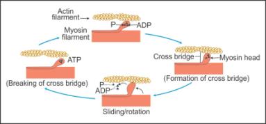
4. Activation of Myosin Heads:
- Because of the release of calcium ions, the myosin heads also get activated and they release energy from ATP.
- Release of energy by the hydrolysis of ATP results in the binding of myosin heads to the active sites present on the actin filaments, forming a cross-bridge.
5. Sliding of Actin Filaments over Myosin:
- As soon as the cross-bridge is formed, the myosin head rotates to pull the actin filament towards the centre of the A-band, i.e. the H-zone.
- The Z-line attached to the actin filaments is also pulled inwards.
- The pulling of actin filaments from the opposite ends results in the contraction of sarcomere.
- During contraction, the I-band shortens, while the A-band retains its length.
- This causes muscle contraction.
Locomotion And Movement Exercise 229
Solution 4
(a) Actin is present in thin filament.
True
(b) H-zone of striated muscle fibre represents both thick and thin filaments.
False
H-zone of striated muscle fibre represents only thick filaments.
(c) Human skeleton has 206 bones.
True
(d) There are 11 pairs of ribs in man.
False
There are 11 pairs of ribs in man.
(e) Sternum is present on the ventral side of the body.
True
Solution 5(a)
|
Actin |
Myosin |
| 1. It forms I-bands (thin filaments) of the myofilament. | 1. It forms A-bands (thick filaments) of the myofilament. |
| 2. It is made of globular actin monomers. |
2. It is made of meromyosin monomers. |
| 3. Regulatory proteins tropomyosin and troponin are associated with the actin. |
3. No regulatory proteins are associated, but each meromyosin is made of two identical heavy chains and four light chains. |
Solution 5(b)
|
Red Muscles |
White Muscles |
|
1. The myoglobin content is high. |
1. The myoglobin content is low. |
|
2. Amount of sarcoplasmic reticulum is moderate. |
2. Amount of sarcoplasmic reticulum is high. |
|
3. Fibres are thin and narrow. |
3. Fibres are thick and broad. |
|
4. Obtain energy from aerobic respiration. |
4. Obtain energy from anaerobic respiration. |
|
5. Red muscles are not fatigued. |
5. White muscles are fatigued. |
|
6. Contain plenty mitochondria. |
6. Contain few mitochondria. |
Solution 5(c)
|
Pectoral Girdle |
Pelvic Girdle |
|
1. It is also called shoulder girdle. |
1. It is also called hip girdle. |
|
2. Each half of the girdle is made of two bones-scapula and clavicle. |
2. Each half of the girdle is made of three bones-ilium, ischium and pubis. |
|
3. It provides articulation to the forelimbs. |
3. It provides articulation to the hindlimbs. |
|
4. The head of the humerus articulates with the glenoid cavity of the pectoral girdle. |
4. The head of the femur articulates with the acetabulum of the pelvic girdle. |
Solution 6
|
Column I |
Column II |
|
(a) Smooth muscle |
(iv) Involuntary |
|
(b) Tropomyosin |
(ii) Thin filament |
|
(c) Red muscle |
(i) Myoglobin |
|
(d) Skull |
(iii) Sutures |
Solution 7
Different types of movements exhibited by the cells of the human body:
1. Amoeboid movements:
- These movements are produced by temporary formation of pseudopodia.
- In protozoans, such as Amoeba, pseudopodia help in capturing food and locomotion.
- Leucocytes and macrophages move in the tissues with the help of pseudopodia.
- Cytoskeletal elements such as microfilaments are also involved in amoeboid movements.
2. Ciliary movements:
- These movements occur in the internal tubular organs which are lined with the ciliated epithelium.
- Cilia lining the trachea propel dust particles out of the body.
- Cilia lining the fallopian tubes and vas efferentia help in the movements of ova and sperms in the desired direction.
- Cilia also help in swimming in ciliated protozoans and ciliated larva such as planula larva of coelenterates and the trochophore larva in annelids.
3. Muscular movements:
- These movements occur in muscle cells which help the organisms in movement and locomotion.
- Muscle cells contract and relax to bring about the movement.
- Movements of limbs, tongue and jaws are examples of muscular movements.
(Note: According to the NCERT textbook, the above-mentioned are movements exhibited by cells. Flagellar movement and cytoplasmic streaming are also movements exhibited by cells).
Solution 8
|
Skeletal Muscles |
Cardiac Muscles |
| 1. They are attached to the primary bones. | 1. They are found in the walls of the heart. |
| 2. Muscle fibres are unbranched. | 2. Muscle fibres are branched. |
| 3. Nuclei are located peripherally. | 3. One nucleus is located at the centre. |
| 4. Intercalated discs are absent in the muscle fibres. | 4. Intercalated discs are present in the muscle fibres. |
| 5. Their function is voluntary. | 5. They function involuntarily. |
| 6. They help in locomotory actions and body posture. | 6. They help in heart movement. |
Solution 9
|
Bones |
Type of Joint Present |
|
(a) Atlas/axis |
Pivot joint |
|
(b) Carpal/metacarpal of thumb |
Saddle joint |
|
(c) Between phalanges |
Hinge joint |
|
(d) Femur/acetabulum |
Ball and socket joint |
|
(e) Between cranial bones |
Fibrous joint |
|
(f) Between pubic bones in the pelvic girdle |
Cartilaginous joint |
Solution 10
- All mammals (except a few) have seven cervical vertebrae.
- The number of phalanges in each limb of human is 14.
- Thin filament of myofibril contains 2 'F' actins and two other proteins namely tropomyosin and troponin.
- In a muscle fibre, Ca++ is stored in sarcoplasmic reticulum.
- 11th and 12th pairs of ribs are called floating ribs.
- The human cranium is made of eight bones.