CBSE Class 10 Answered
How does our heart works? Define in detail.
Asked by | 27 Sep, 2012, 08:55: PM
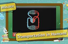
The right-hand side of the heart receives de-oxygenated blood from the body tissues (from the upper- and lower-body via the Superior Vena Cava and the Inferior Vena Cava, respectively) into the right atrium. This de-oxygenated blood passes through the tricuspid valve into the right ventricle. This blood is then pumped under higher pressure from the right ventricle to the lungs via the pulmonary artery.
The left-hand side of the heart receives oxygenated blood from the lungs (via the pulmonary veins) into the left atrium. This oxygenated blood then passes through the bicuspid valve into the left ventricle. It is then pumped to the aorta under greater pressure (as explained below). This higher pressure ensures that the oxygenated blood leaving the heart via the aorta is effectively delivered to other parts of the body via the vascular system of bllod vessels (incl. arteries, arterioles, and capillaries).
The pump action performed by the heart is achieved by a sequence of alternating contraction and relaxation of the heart muscle. This process is directed by the nervous system, nerve impluses initiating each sequence. The whole series of actions that cause alternating contractions and relaxations may be summarised in five stages:
- The vagus nerve stimulates the sinoatrial node (SAN), the pacemaker of the heart.
(The sinoatrial node (SAN) is a tiny area of specialised cardiac muscle in the upper wall of the right atrium, near the vena cava). The fibres of the SAN contract rhythmically approx. 70 times each minute. After each of these contractions, the impluse is dispersed across the atrial cardiac muscle. - This leads to simultaneous contraction of both the right and left atria. This movement of the cardiac muscle pushes blood from the atria into the ventricles (via the tricuspid and bicuspid valves).
- The contractions of the atria send impulses down the Purkinje fibers, which in turn stimulate the atrioventricular node (AVN). The atrioventricular node is a mass of modified cardiac muscle located in the lower/central part of the right atrium of the heart. The Purkinje fibres are a bundle of modified cardiac muscle fibers that transmit impulses from the atra, via the AVN, to the ventricles.
- The action potential from the impulse transmitted down the Purkinje fibers reaches the right and left branches of the Purkinje fibres.
- This causes the ventricles to contract, which pushes blood upwards into the arteries that take the blood away from the heart (the pulmonary artery taking blood to the lungs, and the aorta taking blood to the body).
Answered by | 28 Sep, 2012, 10:50: AM
Application Videos
-
Absorption in small intestine
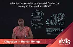 This video explains why absorption of food occurs in the small intestine.
 -
Opening and closing of stomata
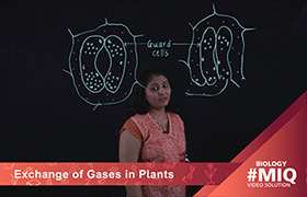 This video explains how guard cells regulate the opening and closing of sto...
 -
Exchange of gases in Amoeba
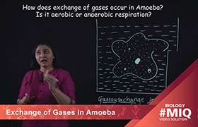 This video details the process of exchange of gases in Amoeba.
 -
Exchange of gases in fishes
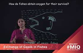 This video details the process of exchange of gases in fishes.
 -
Arteries and Veins
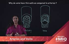 This video explains the difference between arteries and veins.
Concept Videos
-
Respiration in Organisms
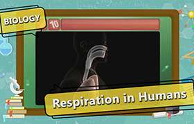 Learn the mechanism of breathing and various organs of respiratory system....
 -
Transport in Organisms
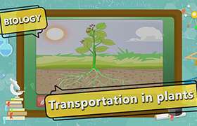 Explain the need for transportation system. Function of the vascular tissu...
 -
Blood
 This video explains the components of blood, blood vessels and the concept ...
 -
Autotrophic Nutrition - Part 1
 This video explains the types of nutrition, photosynthesis in plants and th...
 -
Respiration in Organisms
 Learn the mechanism of breathing and various organs of respiratory system....
CBSE 10 - Biology
Asked by kunchalasrinivasaraosrinivasar | 23 Apr, 2024, 07:40: PM
CBSE 10 - Biology
Asked by bgmigaurav7318 | 08 Apr, 2024, 01:46: PM
CBSE 10 - Biology
Asked by jayeshsah1995 | 04 Mar, 2024, 12:01: PM
CBSE 10 - Biology
Asked by nsubhashree54 | 24 Feb, 2024, 11:08: AM
CBSE 10 - Biology
Asked by sarmabankupalli9 | 02 Jan, 2024, 08:19: PM
CBSE 10 - Biology
Asked by pavanimullu123 | 26 Dec, 2023, 07:17: PM
CBSE 10 - Biology
Asked by jitendranathsingh331 | 26 Nov, 2023, 06:17: PM
CBSE 10 - Biology
Asked by waccstudies123 | 08 Oct, 2023, 02:10: AM
CBSE 10 - Biology
Asked by 09.10bjanvhijadhav | 16 Sep, 2023, 11:58: PM
CBSE 10 - Biology
Asked by devy64469 | 03 Sep, 2023, 09:25: PM